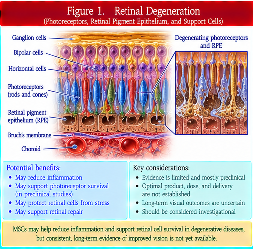
Retinal degeneration is a broad category of disorders in which photoreceptors, retinal pigment epithelium (RPE), or other retinal cells progressively lose function or viability. Etiologies include inherited retinal diseases and acquired degenerative processes, so therapeutic targets differ substantially. MSC and extracellular-vesicle research has explored trophic support, immunomodulation, oxidative-stress reduction, vascular effects, and support of surviving retinal cells. These mechanisms are distinct from true photoreceptor or RPE replacement using retinal progenitor or pluripotent-stem-cell-derived products. Human evidence for MSC/EV therapy remains investigational, and structural or electrophysiologic changes should not be equated automatically with durable visual benefit.
Macular degeneration
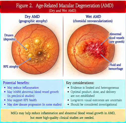
Macular degeneration most commonly refers to age-related macular degeneration (AMD), in which central retinal function is threatened by degeneration of the macula. Nonexudative and neovascular AMD have different biology and established treatment options. Cell-replacement programs have particularly investigated RPE replacement or support, whereas MSC and EV strategies generally emphasize paracrine, anti-inflammatory, trophic, or vascular effects. Evidence from one product, disease stage, or AMD phenotype cannot be generalized to another. Visual acuity, reading function, anatomic outcomes, treatment burden, geographic-atrophy progression, and long-term ocular safety should be distinguished.
Optic-nerve injury
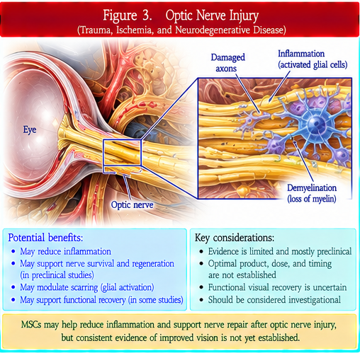
Optic-nerve injury can result from trauma, ischemia, glaucoma, inflammation, compression, or other causes and involves retinal ganglion-cell loss and axonal damage within the optic nerve. MSC and EV research has explored neuroprotection, immunomodulation, trophic signaling, axonal-support mechanisms, and modulation of glial responses. Regenerating a long central nervous system axon and restoring correct synaptic connections is biologically different from transient neuroprotection. Human evidence remains limited, and changes in retinal nerve-fiber-layer thickness, electrophysiology, or isolated visual measures do not establish reliable optic-nerve regeneration.
Corneal disease
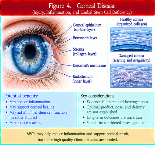
Corneal disease includes epithelial injury, stromal scarring, endothelial dysfunction, limbal stem-cell deficiency, inflammatory disease, infection-related damage, and inherited or degenerative disorders. The cornea is biologically distinct from the retina and optic nerve. Regenerative approaches include established or developing limbal epithelial stem-cell strategies in selected settings as well as investigational MSC, EV, stromal, and endothelial-cell approaches. Outcomes should distinguish epithelial closure, transparency, corneal thickness, endothelial-cell function, neovascularization, pain, visual acuity, graft requirement, and recurrence. Evidence from one corneal layer or disease cannot be generalized across the entire cornea.
Diabetic retinopathy
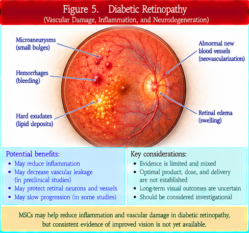
Diabetic retinopathy is a microvascular complication of diabetes involving retinal capillary injury, ischemia, vascular leakage, inflammation, and pathologic neovascularization. Diabetic macular edema and proliferative diabetic retinopathy are important vision-threatening manifestations. MSC and EV research has investigated vascular stabilization, anti-inflammatory signaling, neuroprotection, and tissue support, but angiogenic effects are context-dependent and potentially hazardous in a disease characterized by abnormal neovascularization. Glycemic and blood-pressure management, retinal surveillance, intravitreal anti-VEGF therapy, laser treatment, and vitreoretinal surgery remain established components of care according to disease stage.
Documented risks of unapproved ocular injections
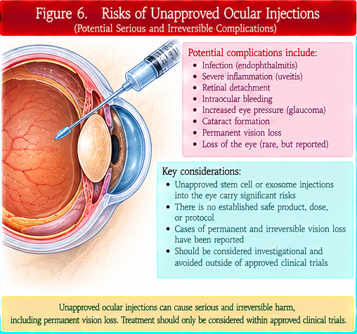
Documented risks of unapproved ocular injections are a central safety issue because the eye is a small, highly specialized organ in which injected products can cause irreversible harm. Reports involving unapproved or poorly characterized cell products have described severe inflammation, retinal detachment, proliferative vitreoretinopathy, hemorrhage, elevated intraocular pressure, infection, cataract, and profound permanent vision loss. Bilateral same-day procedures can magnify catastrophic risk. Marketing claims, payment for participation, trial registration, or use of the term 'stem cell' do not establish product quality, regulatory authorization, or clinical efficacy. Ocular interventions require exact product identity, sterility, dose, route, surgical technique, and long-term ophthalmic follow-up.
Clinical takeaway
Ophthalmic regenerative medicine is anatomically and product specific. Retina, macula, optic nerve, and cornea require different therapeutic strategies, and ocular safety demands unusually strict product and procedural control.
Biological rationale
Proposed mechanisms include trophic support, immunomodulation, neuroprotection, oxidative-stress modulation, vascular stabilization, epithelial repair, and support of retinal or corneal cell survival.
Product identity
MSCs, MSC-derived EVs, retinal progenitors, pluripotent-stem-cell-derived RPE or retinal cells, limbal epithelial stem cells, corneal endothelial cells, and other ocular products are not interchangeable.
Preclinical evidence
Animal and organoid models support multiple retinal, optic-nerve, and corneal hypotheses, but ocular anatomy, disease stage, surgical technique, immune environment, and endpoints affect translation.
Human clinical evidence
Human development varies by indication and product. Some cell-replacement platforms are distinct from MSC/EV therapy; broad class-wide efficacy claims are not justified.
Trial design
Trials should specify eye, laterality, disease phenotype and stage, baseline vision, prior treatment, exact route, sham or active controls when feasible, masked outcome assessment, and long-term structural and functional endpoints.
Dose and administration
Intravitreal, subretinal, suprachoroidal, periocular, topical, intracorneal, and surgically implanted delivery create fundamentally different tissue exposure and procedural risks.
Safety
Risks include endophthalmitis, sterile inflammation, retinal tear or detachment, hemorrhage, proliferative vitreoretinopathy, pressure elevation, cataract, corneal injury, abnormal proliferation, and permanent vision loss.
Regulatory status
Regulatory authorization is exact-product, indication, route, jurisdiction, and date specific. Trial registration, clinic licensure, or payment does not establish authorization or efficacy.
Standard care
Anti-VEGF therapy, laser or vitreoretinal surgery, established corneal transplantation or limbal procedures, glaucoma or inflammatory treatment, metabolic risk control, and low-vision rehabilitation remain central where indicated.
Knowledge gaps
Major gaps include durable cell survival and integration, functional synaptic restoration, immune compatibility, optimal route and dose, potency assays, tumorigenicity surveillance, and long-term ocular safety.
Bottom-line evidence statement
Ophthalmic MSC and EV approaches remain investigational; mechanistic or structural signals are not proof of restored vision, and unapproved ocular injections can cause catastrophic irreversible harm.
Additional controlled terminology
A photoreceptor detects light, while the retinal pigment epithelium supports photoreceptor health. The macula mediates high-resolution central vision; geographic atrophy and neovascular AMD are distinct advanced AMD phenotypes. A retinal ganglion cell sends its axon through the optic nerve, and the retinal nerve fiber layer contains these axons. The corneal epithelium, corneal stroma, and corneal endothelium are distinct layers, and limbal stem-cell deficiency primarily compromises epithelial renewal. Diabetic macular edema and proliferative diabetic retinopathy are vision-threatening diabetic complications involving leakage or neovascularization. Anti-VEGF therapy is established for several neovascular retinal conditions. Intravitreal injection and subretinal delivery are different routes. Endophthalmitis, retinal detachment, proliferative vitreoretinopathy, and elevated intraocular pressure can threaten vision. Visual acuity is an important but incomplete measure of visual function.