Plain-language overview
Ophthalmology includes several very different tissues and diseases. The retina, macula, optic nerve, and cornea have different structures and functions, so a treatment studied for one should not be assumed to work for another.
Research involving mesenchymal stromal cells (MSCs), extracellular vesicles (EVs), and other cell products has examined tissue support, inflammation, nerve protection, vascular effects, and true cell-replacement strategies. These approaches are not interchangeable.
Because vision can be permanently damaged by an unsafe procedure, the exact product, route, dose, disease, and clinical evidence matter greatly. Unapproved or poorly characterized ocular injections have caused severe irreversible injury.
Retinal degeneration
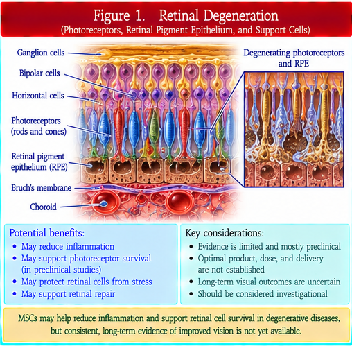
Retinal degeneration is a broad group of disorders in which photoreceptors, retinal pigment epithelium, or other retinal cells lose function or die.
A photoreceptor detects light. The retinal pigment epithelium supports photoreceptor health and performs several functions needed for normal retinal activity.
MSC and EV research has examined protective, anti-inflammatory, and tissue-supportive effects. This is different from actually replacing retinal cells with retinal progenitors or cells derived from pluripotent stem cells. Changes in retinal structure or electrical testing do not automatically prove durable improvement in vision.
Macular degeneration
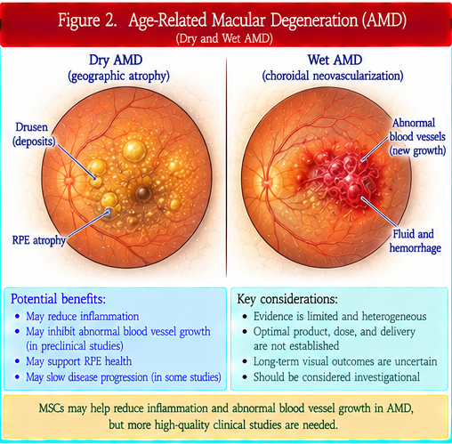
Macular degeneration most commonly refers to age-related macular degeneration. The macula is the part of the retina responsible for detailed central vision.
Geographic atrophy and neovascular AMD are different advanced forms of the disease and require different treatment strategies.
Some research programs aim to replace or support retinal pigment epithelial cells, while MSC and EV approaches generally investigate supportive or signaling effects. Evidence from one product or form of AMD should not be generalized to another. Visual acuity, reading ability, anatomy, treatment burden, disease progression, and long-term safety are separate outcomes.
Optic-nerve injury
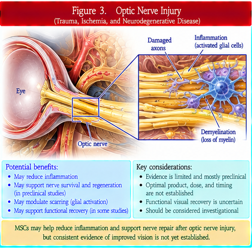
Optic-nerve injury can result from trauma, poor blood flow, glaucoma, inflammation, compression, and other causes.
A retinal ganglion cell sends an axon through the optic nerve. The retinal nerve fiber layer contains these axons within the retina.
MSC and EV research has explored nerve protection, immune regulation, and trophic signaling. Protecting a surviving nerve cell is very different from regenerating a long damaged axon and reconnecting it correctly in the brain. Current human evidence does not establish reliable optic-nerve regeneration.
Corneal disease
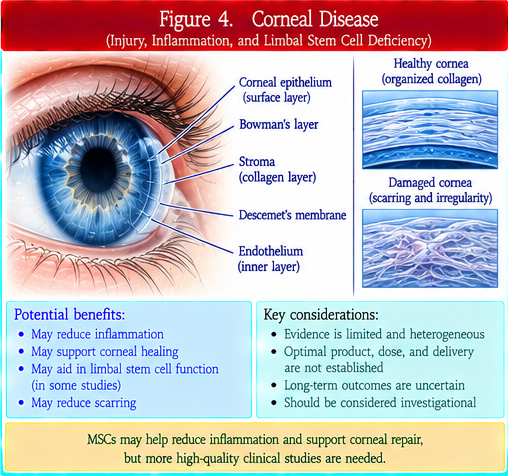
Corneal disease can involve several different layers. The corneal epithelium forms the outer surface, the corneal stroma provides most of the cornea’s thickness and transparency, and the corneal endothelium helps maintain appropriate hydration.
Limbal stem-cell deficiency interferes with normal renewal of the corneal surface. Selected limbal epithelial stem-cell strategies are distinct from investigational MSC, EV, stromal-cell, or endothelial-cell approaches.
Meaningful outcomes include healing of the surface, transparency, corneal thickness, endothelial function, abnormal blood-vessel growth, pain, visual acuity, need for transplantation, and recurrence.
Diabetic retinopathy
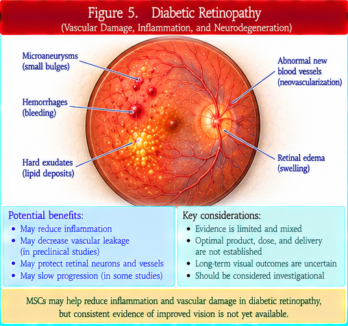
Diabetic retinopathy is a microvascular complication of diabetes in which retinal blood vessels can become damaged, leaky, ischemic, or abnormally proliferative.
Diabetic macular edema involves fluid accumulation affecting the macula. Proliferative diabetic retinopathy involves pathologic neovascularization.
MSC and EV research has examined vascular stabilization, inflammation, nerve protection, and tissue support. However, stimulating blood-vessel growth can be hazardous in a disease already characterized by abnormal vessels. Anti-VEGF therapy, metabolic risk control, laser treatment, and vitreoretinal surgery remain established treatments when appropriate.
Documented risks of unapproved ocular injections
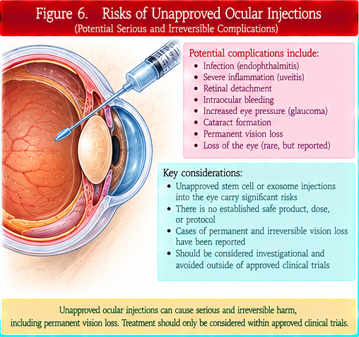
Unapproved ocular injection is a major safety concern because injected material can directly damage delicate structures needed for vision.
Documented complications of unapproved or poorly characterized cell injections include severe inflammation, endophthalmitis, retinal detachment, proliferative vitreoretinopathy, hemorrhage, increased intraocular pressure, cataract, infection, and profound permanent vision loss.
An intravitreal injection places material into the vitreous cavity, while subretinal delivery places material beneath the retina. These and other ocular routes have different risks. Trial registration, clinic licensure, payment for participation, or use of the words “stem cell” does not prove that a product is authorized, safe, or effective.
Why ocular products and routes differ
MSCs, MSC-derived EVs, retinal progenitor cells, retinal pigment epithelial cells, limbal epithelial stem cells, corneal endothelial cells, and other ocular cell products are biologically different interventions.
Intravitreal, subretinal, suprachoroidal, periocular, topical, intracorneal, and surgically implanted delivery expose different tissues and create different procedural risks.
How ophthalmology studies can mislead
Imaging, retinal thickness, electrophysiology, cell survival, or other structural measurements can provide useful research information without proving that vision has meaningfully improved.
Visual acuity is important but does not capture every aspect of visual function. Strong studies should also consider disease-specific functional outcomes, durability, anatomy, adverse events, and treatment burden.
Safety considerations
Ocular procedures can cause infection, sterile inflammation, retinal tears or detachment, bleeding, abnormal scar formation, pressure elevation, cataract, corneal injury, and permanent vision loss.
Cell products may add additional concerns involving immune reactions, abnormal proliferation, manufacturing quality, sterility, or long-term survival. Bilateral same-day procedures can expose both eyes to the same catastrophic risk.
Questions patients should ask
What exact eye disease and structure are being treated? What exact cell or EV product is being used? Is the goal tissue support or true cell replacement? What route will be used? Is the product authorized for this exact indication? What controlled human evidence shows meaningful visual benefit? What serious ocular complications have occurred? Are both eyes being treated at the same time? What established ophthalmic treatments are available?
Bottom line
Ophthalmic regenerative research is active, but the evidence is highly specific to the eye structure, disease, product, and route.
Mechanistic or structural changes are not proof that vision has been restored. Unapproved ocular cell injections deserve particular caution because severe, irreversible vision loss has been documented.
How this review was prepared
This Patient Edition was derived directly from the explicitly approved 0810 Professional Edition and preserves all six controlling ophthalmology Points.