SCE Educational Library
6. Exosomes and Other Extracellular Vesicles
6.8 EV characterization
Particle concentration
Particle size
Electron microscopy
Flow cytometry
Western blotting
Proteomics
Lipidomics
RNA analysis
Single-vesicle analysis
Plain-language overview
Extracellular-vesicle characterization is the process of measuring what is present in an EV preparation and what can reasonably be concluded from those measurements. No single test can prove EV identity, purity, exosome origin, biological potency, safety, or clinical effectiveness.
Strong characterization therefore uses several different types of evidence together. Researchers may measure how many particles are present, their size, their appearance, their surface markers, their protein and lipid content, their RNA cargo, and—increasingly—the properties of individual vesicles rather than only averages across the entire sample.
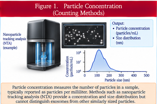
Particle concentration
Particle concentration estimates how many detectable particles are present in a defined volume. Common methods include nanoparticle tracking analysis and tunable resistive-pulse sensing.
The reported number depends on the instrument, detection threshold, dilution, background particles, refractive properties, and sample preparation. A particle count does not tell us how many detected particles are actually EVs, how many belong to a particular EV subtype, or how many carry a proposed active cargo.
Particle size
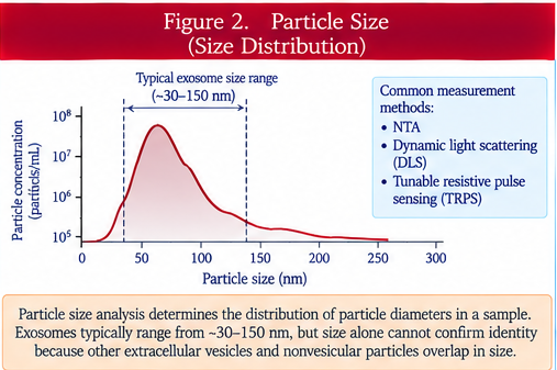
Particle size describes the physical dimensions or size distribution of particles detected in the sample. Methods such as nanoparticle tracking analysis, dynamic light scattering, resistive-pulse sensing, and electron microscopy can provide size information.
Different instruments measure different physical properties and have different biases. EVs also overlap in size with lipoproteins, protein aggregates, viruses, and other nanoparticles, so size alone cannot prove that a particle is an exosome or even an EV.
Electron microscopy
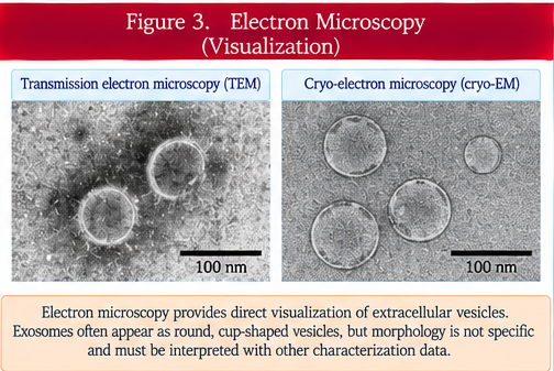
Electron microscopy provides direct structural images of very small particles. Transmission electron microscopy and cryogenic electron microscopy are commonly used approaches.
These techniques can show membrane-bound particles and their morphology, but sample preparation can alter appearance and the field of view usually represents only a small fraction of the entire preparation. An electron-microscopy image therefore supports structural characterization but does not establish purity or biological identity by itself.
Flow cytometry
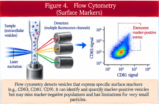
Flow cytometry can detect particles carrying fluorescent labels or selected surface molecules. Specialized instruments and careful calibration are required because EVs are much smaller and dimmer than most cells.
Small-particle measurements are vulnerable to swarm detection and coincidence, in which multiple particles are counted as one event. Refractive index, instrument settings, reference particles, fluorescence-minus-one controls, detergent sensitivity, and appropriate blanks all help determine whether a signal is trustworthy.
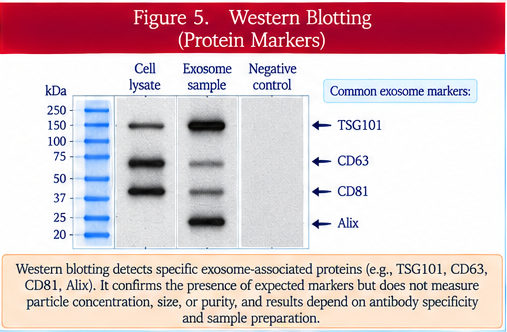
Western blotting
Western blotting detects selected proteins in a bulk EV preparation. Researchers may test for positive markers expected to be enriched in EVs and negative markers that help identify unwanted cellular contamination.
A positive marker does not mean every particle carries that protein, and a negative marker does not prove complete absence of contaminants. Western blotting is most useful as one part of a broader characterization panel.
Proteomics
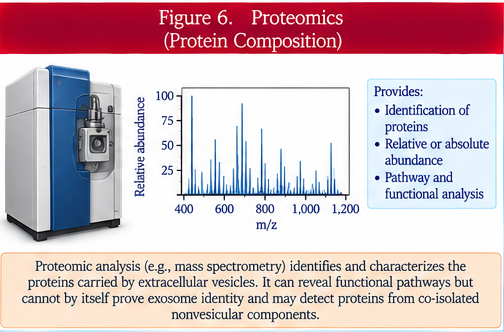
Proteomics studies the protein composition of a preparation, usually with mass spectrometry and peptide identification. It can reveal hundreds or thousands of proteins and help compare products, cell sources, or manufacturing conditions.
Because proteomics measures the bulk sample, detected proteins may come from EVs, co-isolated proteins, or other particles. The results therefore need to be interpreted together with purity measurements and complementary assays.
Lipidomics
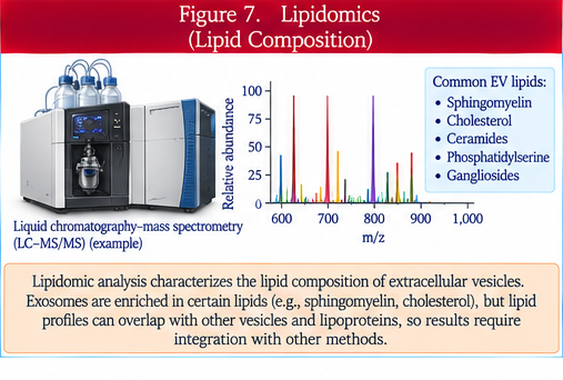
Lipidomics examines the lipid composition of EV preparations. EV membranes contain complex mixtures of phospholipids, cholesterol, sphingolipids, and other lipids that can influence membrane structure and biological interactions.
Lipid profiles can help compare EV populations and manufacturing conditions, but bulk lipid measurements do not automatically show which lipids are present on individual vesicles or whether the measured lipids are responsible for a biological effect.
RNA analysis
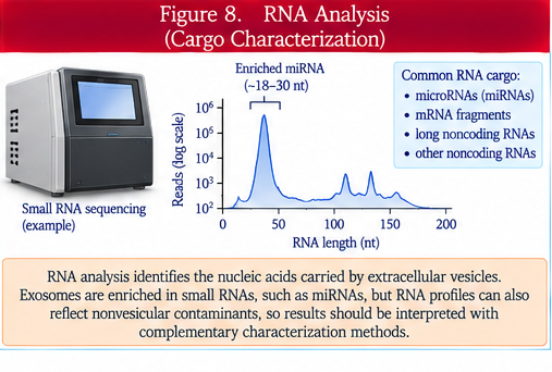
EV preparations can contain messenger RNA, microRNA, and other RNA species. RNA analysis may use digital PCR, RNA sequencing, and RNase protection experiments.
These methods can show that RNA is associated with a preparation, but they do not automatically prove that the RNA is inside EVs, present in every vesicle, delivered to recipient cells, or biologically active. RNase protection and other controls help distinguish protected vesicular RNA from external RNA contamination.
Single-vesicle analysis
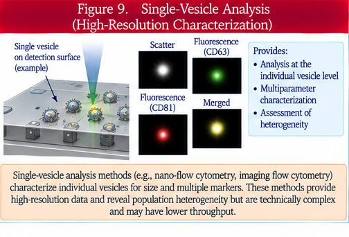
Single-vesicle analysis examines individual particles rather than reporting only an average for the entire preparation. These methods can reveal heterogeneity that bulk measurements may hide.
The challenge is that many instruments detect only a subset of vesicles within a limited size or fluorescence range. Single-vesicle methods are therefore powerful but still require careful calibration, controls, and interpretation.
Why several methods are needed
Different characterization methods answer different questions. Particle counting measures abundance, microscopy measures structure, flow cytometry and western blotting assess selected markers, proteomics and lipidomics examine composition, and RNA analysis measures nucleic-acid content. Combining independent methods provides stronger evidence than relying on one assay.
Controls and reference materials
Reliable measurements require appropriate blanks, reagent controls, dilution studies, reference particles, calibration standards, spike-recovery testing, and predefined acceptance criteria. The controls must match both the assay and the sample being tested.
Bulk measurements versus single-particle measurements
Bulk assays report the average composition of the whole preparation and can hide important subpopulations. Single-vesicle analysis can expose heterogeneity but may not detect every vesicle. Both approaches can therefore provide useful but different information.
Why manufacturing matters
For a clinical product, characterization assays should be qualified or validated for their intended purpose. They may be used for lot release, stability testing, comparability after manufacturing changes, or process control.
Questions patients should ask
Which characterization tests were performed? What did each test actually demonstrate? Were contaminants measured? Were appropriate controls and calibration standards used? Was the product tested across multiple manufacturing lots? Was endosomal origin directly demonstrated, or was the word exosome inferred from size or a small marker panel?
Bottom line
EV characterization is a multidimensional evidence package, not a single test result. Strong characterization combines particle measurements, structural imaging, molecular markers, protein and lipid composition, RNA analysis, contaminant assessment, and increasingly single-vesicle information.
The safest interpretation is to describe only what the assays directly show. A particle count, size range, marker result, microscopy image, or molecular profile should not be stretched into claims of purity, potency, exosome identity, safety, or clinical effectiveness without additional evidence.
How this review was prepared