SCE Educational Library
6. Exosomes and Other Extracellular Vesicles
6.7 EV separation methods
Differential centrifugation
Ultracentrifugation
Density gradients
Size-exclusion chromatography
Ultrafiltration
Tangential-flow filtration
Precipitation
Immunoaffinity capture
Plain-language overview
Extracellular-vesicle, or EV, separation methods are ways of enriching vesicles from fluids that also contain cells, proteins, lipoproteins, debris, and many other particles. No method produces a perfectly pure EV population by itself, and no separation method alone proves that the particles are exosomes.
Each method separates material according to a practical property such as size, density, sedimentation, membrane passage, solubility, or binding to a selected surface molecule. Because different methods enrich different mixtures, the method used can change the measured composition, yield, purity, and biological activity of the final preparation.
Differential centrifugation
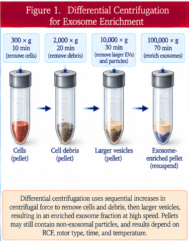
Differential centrifugation uses a series of spins at progressively higher centrifugal forces. Lower-speed spins remove intact cells and larger debris first; higher-speed spins then enrich smaller particles.
This approach is useful for clarification and preliminary enrichment, but the resulting pellets can contain a mixture of EVs, protein aggregates, organelles, and other material. The actual relative centrifugal force, spin time, rotor type, temperature, and sample handling all affect the result.
Ultracentrifugation
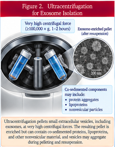
Ultracentrifugation uses very high centrifugal forces to sediment small extracellular particles. It remains common in EV research because it does not require a binding reagent and can process relatively large sample volumes.
However, proteins, lipoproteins, and other nanoparticles can co-sediment with EVs. High-force pelleting can also promote aggregation and make the pellet difficult to resuspend. A pellet obtained this way should therefore be described as an enriched preparation unless additional testing demonstrates greater purity.
Density gradients
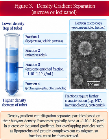
Density-gradient methods separate particles according to buoyant density. The sample is placed in or over a gradient, commonly made with iodixanol or sucrose, and centrifuged until particles move to regions matching their density.
Density gradients can improve separation from some protein aggregates and other contaminants, but overlapping densities still occur. The method is labor-intensive and recovery can be incomplete, so fractions need additional characterization rather than being assumed to contain one pure EV subtype.
Size-exclusion chromatography
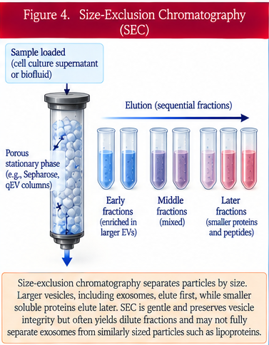
Size-exclusion chromatography, or SEC, passes a sample through a column containing porous beads. Larger particles such as many EVs move around the beads and come out earlier, while smaller soluble proteins enter the pores and are delayed.
SEC can preserve vesicle structure and often separates EVs from much of the soluble protein content. It does not, however, distinguish EVs from every other particle of similar size, and the early fractions can still contain lipoproteins or other nanoparticles.
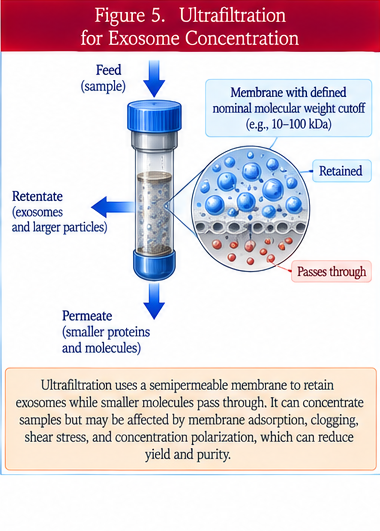
Ultrafiltration
Ultrafiltration uses a membrane with a defined pore size or molecular-weight cutoff to retain larger particles while smaller molecules pass through. It can be used to concentrate EV-containing samples or remove smaller solutes.
Recovery depends on membrane material, pore characteristics, pressure, sample concentration, and how much material sticks to the filter. Very high pressure or excessive concentration can alter recovery or damage vesicles.
Tangential-flow filtration
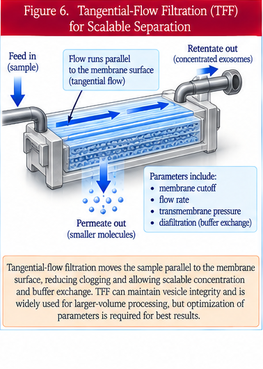
Tangential-flow filtration, or TFF, is a form of membrane filtration in which fluid moves parallel to the membrane surface rather than directly into it. This reduces rapid clogging and allows larger volumes to be concentrated and washed.
TFF can be scalable and useful for manufacturing, but performance depends on membrane choice, transmembrane pressure, flow rate, diafiltration strategy, and system design. It enriches particles by size-related filtration and does not by itself establish EV identity or purity.
Precipitation
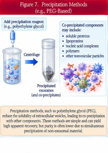
Precipitation methods add substances such as polyethylene glycol that reduce particle solubility and cause extracellular particles to collect together so they can be recovered.
These methods are simple and can produce high apparent recovery, but they can also co-precipitate proteins, lipoproteins, nucleic acids, and other nonvesicular material. High particle yield therefore should not be confused with high EV purity.
Immunoaffinity capture
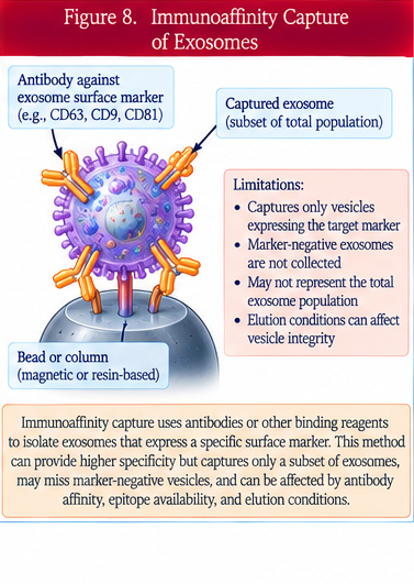
Immunoaffinity capture uses antibodies or other binding molecules to isolate particles carrying a selected surface epitope. For example, a capture reagent may bind a tetraspanin or another EV-associated surface protein.
This approach can enrich a defined subpopulation, but it only captures particles that display the chosen target and may miss other EVs. The selected marker is not necessarily unique to one EV subtype, and strong binding can make recovery of intact vesicles difficult.
Why no single method is universally best
Every separation method creates tradeoffs among yield, purity, scalability, speed, cost, equipment requirements, and preservation of vesicle function. A method that is useful for discovery research may not be ideal for clinical manufacturing, and a high-yield method may produce a less pure preparation.
Why characterization must follow separation
Separation is only the first step. The resulting preparation should be characterized for particle concentration, size distribution, protein and lipid markers, contaminants, morphology when appropriate, and other quality attributes relevant to the intended use. A separation method does not substitute for product characterization.
What patients should understand about product claims
Terms such as exosome preparation, purified EVs, or high-purity exosomes should be supported by data rather than inferred from the name of the separation method. Patients and clinicians should ask how the product was separated, what contaminants were measured, how identity was assessed, whether lots are consistent, and whether the final preparation was tested for potency and safety.
Questions patients should ask
Which separation method was used? Was more than one method combined? What was the recovery and purity? Were proteins, lipoproteins, nucleic acids, and other contaminants measured? How were particle identity and concentration determined? Were the same methods used for every manufacturing lot? Is there evidence that the separation process preserves the proposed biological activity?
Bottom line
EV separation methods enrich extracellular particles according to physical or biochemical properties such as sedimentation, density, size, membrane passage, precipitation behavior, or surface binding. None of these approaches alone proves EV identity, purity, or exosome origin.
The best interpretation considers the full workflow: sample source, preprocessing, separation method, recovery, contaminants, characterization, potency, lot consistency, storage, safety, and the evidence supporting the intended clinical use.
How this review was prepared